| FishBase |
Larvae Information Summary for
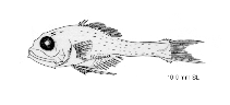
Poromitra megalops
| Main Ref: | Sandknop, E.M. and W. Watson 1996 | |||
Yolk-sac larvae
| max | min | mod | Ref. | |
| Length at birth (mm) | 2.4 | |||
| Preanal L. % TL |
| Place of development | planktonic | |||
| Larval area | Northeastern pacific (California Current region); Northwest Pacific (Japan) | |||
| Yolk-sac | Ref: | |||
| Yolk | Oil globules | |||
| Rows on tail | ||||
| Other melanophores on tail | ||||
Post larvae
| Striking feature | none | |||
| Striking shape lateral | normal (not striking) | dorsal | ||
| Striking feature | none | |||
| Shape of gut | triangular | |||
| Gas bladder early | pigmented | late | ||
| Spinal armature early | opercular spines only | late | several different spines | |
| Pigmentation early | ||||
| Rows on tail | ventral row | |||
| Other melanophores on tail | no other melanophores | |||
| Melanophores on head + trunk | melanophores on head | |||
| Rows on tail | lateral row | |||
| Other melanophores on tail | tail partly covered with melanophores | |||
| Melanophores on head + trunk | melanophores on head + trunk | |||
| Pectorals | normal without melanophores | |||
| Pelvics | normal (i.e. small or absent) without melanophores | |||
| Myomeres 7-10+18-21=27-29 (usually 7+21 in preflexion stage, 9+19 by postflexion stage); preopercular and opercular spines form at end of flexion stage; posttemporal spines form during mid-postflexion stage (about 8 mm); frontal and nasal spines form late in postflexion stage; eyes relatively large; longitudinally elongate dash-like melanophores on trunk and tail beginning in late flexion or early postflexion stage; little or no pigment on pectoral and pelvic fins. Pigmentation: Preflexion: under mid- and hindbrain; along cleithrum; on gas bladder and upper part of gut; few along ventral margin of tail. Flexion: dorsolaterally posteriorly on midbrain, around lower half of forebrain; internally near posterior margin of eye after 6 mm; longitudinally elongate dashes, primarily near dorsal and ventral margins and along lateral midline initially, filling in laterally from trunk to tail; few on caudal fin. Postflexion: on frontal above eye; over posterior part of midbrain between 6.4-11.5 mm; proximal bar across principal caudal fin rays by 11.8 mm. Sequence of fin development: dorsal and pectorals and pelvics, caudal, anal. | ||||
Meristic characters
| max | min | mod | Ref. | |
| Total number of myomeres | 29 | 27 | 36655 |
| L 1st feeding | Ref. | Months of presence of larvae | ||||
| max | Jan | Feb | Mar | Apr | ||
| min | May | Jun | Jul | Aug | ||
| mod | Sep | Oct | Nov | Dec | ||
| Water parameters Metric characters |
| Back to Search |