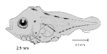
| Relatively deep-bodied; terminal gut section curves anteriad in flexion stage yielding short preanal distance; 4 distinct dorsal melanophores in preflexion larvae; strong preopercular spination, a maximum of 6-7 above and 8-10 below angle spine in posterior series and 3-4 upper and 9-11 lower spines in anterior series; multiple spines on supraocular ridge, posttemporal and supracleithrum; pronounced, scalloped supraoccipital crest.
Pigmentation:
Preflexion: By 2.2 mm, 0-1 medially posteriorly above midbrain, at angular, isthmus, below pectoral fin base, on ventral margin of gut, distally on preanal finfold, above gas bladder and terminal section of gut, 11-13 on ventral margin of tail (smaller posteriorly), 4 patches on dorsal midline; by 3.0 mm, on tip of lower jaw, pair on snout (usually), greater or equal to 1 laterally on jaw, 1-3 on lateral midline anteriorly on tail; by end of stage, several posteriorly above midbrain and anteriorly above hindbrain, medially on gular membrane, 3-6 in lateral midline series, series above spinal column medial to lateral midline series.
Flexion: Above forebrain; 0-1 on opercle; 2 rows on postanal ventral margin; series forming on ventral margin of anal fin base; forming on hypaxial myosepta of tail.
Postflexion: By 7.0 mm, more above brain and on snout, series on each side of dorsal fin base, forming in myosepta above lateral midline streak, laterally on abdomen, on lower hypural region; by 9.0 mm, brain covered, myoseptal zone expanding, solid sheath forming on nape and shoulder region, a blotch forming anteriorly on anal fin; by 11.0 mm, dorsal half of head, and body nearly covered, dorsal fin and anal fin pterygiophores outlined, and distally on dorsal fin, anal fin, and caudal fin.
Juvenile: Late postflexion stage pattern augmented.
Sequence of fin development: principal caudal fin rays, dorsal fin and anal fin and pectoral fin, procurrent caudal fin rays, pelvic fin. |
