FishBase
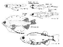
Larvae Information Summary for  Loweina rara
Show available picture(s) for

Main Ref: Moser, H.G. and E.H. Ahlstrom 1996
Yolk-sac larvae
  max min mod Ref.
Length at birth (mm) 2.8      
Preanal L. % TL 81 54    
Place of development planktonic
Larval area California Current region
Yolk-sac       Ref:  
Yolk   Oil globules  
Rows on tail  
Other melanophores on tail  
Both dorsal and ventral fin folds develop into extremely large inflated and translucent sinuses which act as a hydrostatic organ. The main function of this organ is to take up surplus water from the absorbed yolk when that water cannot be excreted through membranes, blood vessels, or excretory organs. Water can be expelled from the fin-fold sinus when it has fully developed (Ref. 26281).
Post larvae
Striking feature light organs
Striking shape lateral normal (not striking) dorsal  
Striking feature light organs
Shape of gut elongated
Gas bladder   early   late  
Spinal armature   early no spines late no spines
Pigmentation early
Rows on tail no rows
Other melanophores on tail no other melanophores
Melanophores on head + trunk melanophores on head + trunk
Pigmentation late
Rows on tail no rows
Other melanophores on tail no other melanophores
Melanophores on head + trunk melanophores on head + trunk
Urostyle region   early unpigmented late unpigmented
Pectorals other melanophores at fin base
Pelvics fin rays only without melanophores
Among largest myctophid larvae at transformation (>20.0 mm SL); early larvae slender but relative body depth doubles in later stages; only Br2 develops during larval period; distinguished from all other myctophids except Tarletonbeania crenularis by: enlarged median folds; distinguished from T. crenularis by: eye moderately narrow, no choroid tissue on ventral surface of eye; band on head between eyes. Compressed body with voluminous dorsal and ventral finfolds; large head; relatively wide oval eyes; elongate gut with large terminal section; large pectoral fins with elongate ornamented lower ray; dorsal and anal fins far posteriad; distinctive pigment pattern, including transverse bar between fore- and midbrain (lacking in Tarletonbeania crenularis). Pigment: Preflexion: by 4.0 mm, a heavy transverse bar between fore- and midbrain, an embedded blotch anterior to pectoral fin base, a blotch embedded above midgut, a blotch on dorsal surface of terminal section of gut, apposing dorsal and ventral blotches in mid-postanal region, and another more anterior dorsal blotch; by 6.0 mm, a median blotch embedded in isthmus, an embedded melanophore at nape, and a blotch on spatulate swellings of elongate lower pectoral fin ray; by end of stage, embedded blotches at pectoral fin base and isthmus expanded toward each other to outline ventral border of gill cavity and postanal blotches lost in some specimens. Postflexion: numerous (large) blotches in voluminous finfold; several embedded in postanal hypaxial myosepta; several on pectoral fin base. Sequence of fin development: pectorals, caudal, anal, dorsal, pelvics.
Meristic characters
  max min mod Ref.
Total number of vertebrae 39 37   31442
  L 1st feeding Ref. Months of presence of larvae
max     Jan Feb Mar Apr
min   May Jun Jul Aug
mod     Sep Oct Nov Dec
Water parameters     Metric characters
Back to Search
Comments & Corrections

php script by kbanasihan, 09/07/10 ,  last modified by sortiz, 09/08/17